New Graphene-Based Material Developed for Medical Implants
- News
- 2K
A group of scientists has developed a new material for biomedical applications by combining a graphene-based nanomaterial with Hydroxyapatite (HAp), a commonly used bioceramic in implants.
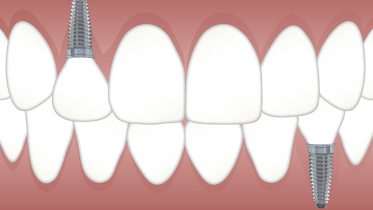
In recent years, biometallic implants have become popular as a means to repair, restructure or replace damaged or diseased parts in orthopedic and dental procedures. Metal parts also find use in devices such as pacemakers.
However, metallic implants face several limitations and are not a permanent solution. They react with body fluids and corrode, release wear and tear debris resulting in toxins and inflammation. They also have high thermal expansion and low compressive strength causing pain and are dense and may cause reactions.
On the other hand, bioceramics do not have these limitations. HAp specifically is osteoconductive, with a bone-like porous structure offering the required scaffold for tissue re-growth. However, it is brittle and lacks the mechanical strength of metals. The problem is overcome by combining it with nanoparticles of materials such as Zirconia.
In the new research, scientists have combined HAp with graphene nanoplatelets. “Previously reported studies have focused on only structural properties of such composites without throwing light on their biological properties. We have found that combining HAp with graphene nanomaterial enhances mechanical strength, provides better in-vivo imaging and biocompatibility without changing its basic bone-like properties,” explained Dr. Gautam Chandkiram, the principal investigator at the University of Lucknow, while speaking to India Science Wire.
Purification of the base ceramic material is a significant primary challenge in fabricating composites. According to scientists, in the current study, highly efficient biocompatible Hydroxyapatite was successfully prepared via a microwave irradiation technique and the consequent composites were synthesized using a simple solid-state reaction method.
The process involved mixing different concentrations of graphene nanoplatelet powders and drying, crushing, sieving and ball-milling the resulting slurry. The fine composite powder was further cold-compressed and sintered at 1200 degrees Celsius to achieve the desired density.
The scientists found that the composite had an adequate interfacial area between the nanoparticles, with the graphene nanoplatelets well distributed into the hydroxyapatite matrix, while exhibiting high fracture resistance. Further, structural characterization, mechanical and load bearing tests showed that the 2D nature of graphene improves the load transfer efficiency significantly.
Researchers also examined cell viability of the composite by observing metabolic activity in specific cells using a procedure known as MTT assay. They used gut tissues of Drosophila larvae and primary osteoblast cells of a rat. “The overall cell viability studies demonstrated that there is no cytotoxic effect of the composites on any cell type,” explained Dr. Gautam.
Biomaterials also find use in drug delivery and bioimaging diagnosis. “Our research on the composite found that it displays a better fluorescence behavior as compared to pure hydroxyapatite, indicating it has a great potential in bone engineering and bioimaging bio-imaging applications as well,” he added.
The research team included Sunil Kumar and Gautam Chandkiram (Advanced Glass and Glass Ceramics Research Laboratory, University of Lucknow); Vijay Kumar Mishra and Ritu Trivedi (CSIR-Central Drug Research Institute, Lucknow); Brijesh Singh Chauhan, Saripella Srikrishna, Ram Sagar Yadav and Shyam Bahadur Rai (Banaras Hindu University). The study results have been published in the journal ACS Omega. (India Science Wire)
If you liked this article, then please subscribe to our YouTube Channel for the latest Science & Tech news. You can also find us on Twitter & Facebook.


