Scientists Decipher How Japanese Encephalitis Virus Enters Brain Cells
- News
- 2.5K
Japanese Encephalitis, a leading form of viral encephalitis is a major problem in India. It is caused by mosquito-borne JE virus which belongs to the same genus as dengue and yellow fever.
In a new study, a team of researchers led by scientists of the National Brain Research Centre (NBRC) has identified proteins that facilitate entry of JE virus inside the brain cells. Two brain cell proteins – PLVAP (Plasmalemma vesicle-associated protein) and GKN3 (Gastrokine3) – facilitate the entry of the virus into the brain cell. The study has been done in laboratory mice.
JE infection occurs when viral attachment proteins interact with cellular membrane proteins of host cells. In their study, scientists synthesized JEV E-glycoprotein (viral attachment protein) in bacteria followed by its collection and purification. The purified E protein was made to interact with cell membrane fraction of mouse brain to find out interacting partners from the cell membrane.

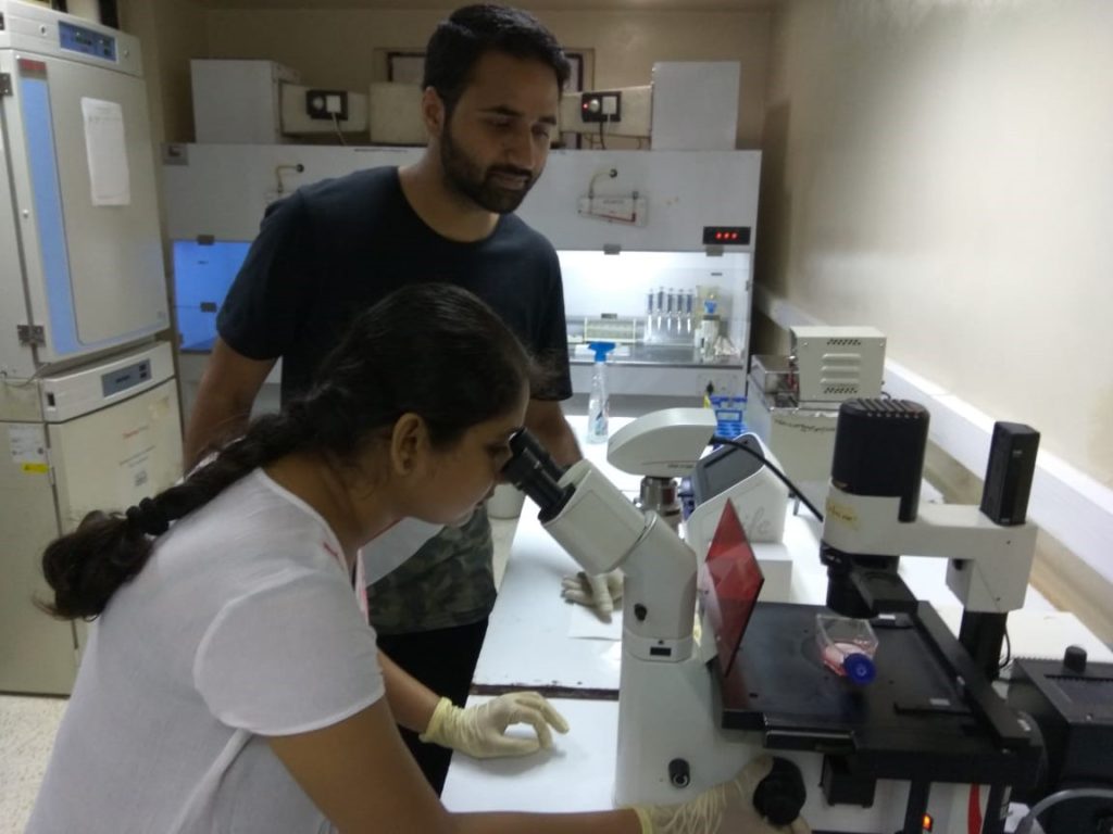
Dr. Anirban Basu with his team.
Following the binding of E protein with cellular partners, the interacting proteins were identified using two-dimensional gel electrophoresis and mass spectrometry.
“Amongst the identified proteins, PLVAP and GKN3 were shown to be present on the neuronal cell surface which means they have a role in viral entry into neurons. We reconfirmed their presence on the surface of neurons isolated from mouse cerebral cortex, which validates their identity as neuronal proteins. In addition, PLVAP protein was found to be upregulated in human autopsy tissue of JE infection,” explained Dr. Anirban Basu, senior scientist at NBRC who led the study.

Team member Sriparna Mukherjee, and Irshad Akbar engaged in research work.
Researchers also found that reducing PLVAP receptor in neurons decreased JEV entry and upregulating (increasing) them increased viral entry on the other hand. It shows the two proteins are important for viral invasion. “Identification of host proteins, important for viral entry into neurons provides us with candidates, whose targeting may be useful to block viral infection thus reducing disease severity,” added Dr. Basu.
Now the team is planning to collaborate with some pharmaceutical companies to identify the potential drug which may be able to block these receptor proteins. According to Dr. Basu, these receptors may be common for other viruses which target cells of the nervous system, like Zika, West Nile, and even dengue. However, extensive research is needed for the confirmation.
“This research is highly significant and will advance the field towards the development of therapies against JE”, commented Dr. Manjula Kalia, a scientist from Faridabad-based Translational Health Science And Technology Institute, who is not associated with this study.
Dr. Milind Gore, a former scientist from National Institute of Virology, Pune, said: “this is one step further in understanding how 10-day old mice are sensitive for JE multiplication and hopefully some good work will come out in future”.
The research team included Dr. Anirban Basu, Sriparna Mukherjee, Nabonita Sengupta, Irshad Akbar, Noopur Singh (National Brain Research Centre, Manesar); Amol Ratnakar Suryawanshi (Institute of Life Sciences, Bhubaneswar); Ankur Chaudhuri and Sibani Chakraborty (West Bengal State University); and Arindam Bhattacharyya (University of Calcutta).
The study has been published in journal Scientific Reports. The research was funded by Department of Biotechnology (DBT) and Council of Scientific and Industrial Research (CSIR). (India Science Wire)
By Yogesh Sharma
Journal Reference
If you liked this article, then please subscribe to our YouTube Channel for the latest Science and Tech news. You can also find us on Twitter and Facebook.


